incisive canal radiograph
Periapical radiograph panoramic and CBCT are needed to assess the lesion with better precision and limits radiation. Mean sd vertical diameter buccolingual diameter and inner diameter of the incisive canal were 47 11 37 07 and 11 03 mm respectively.

Visibility Of Mandibular Anatomical Landmarks In Panoramic Radiography A Retrospective Study Semantic Scholar
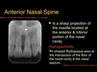
It is located in the maxilla in the incisive fossa midline in the palate posterior to the central incisors at the junction of the medial palatine and incisive sutures.

. Usually only the inferior border of the orbit is visible over the panoramic radiograph Incisive canal. However complications may arise due to an extension anterior to the mental foramen that forms the mandible incisive canal MIC. Today panoramic radiographs OPGs are routinely used in the dental office for various diagnostic purposes that.
Die Bewertungen umfassten 1 den mesiodistalen Durchmesser 2 den labiopalatalen Durchmesser 3 die Länge des Schneidekanals 4 die Form des Schneidekanals und 5 die Breite des Knochens vor dem Foramen incisive. Original uploader was DRosenbach at enwikipedia. An incisive canal was identified in 15 of the images with good visibility in only 1.
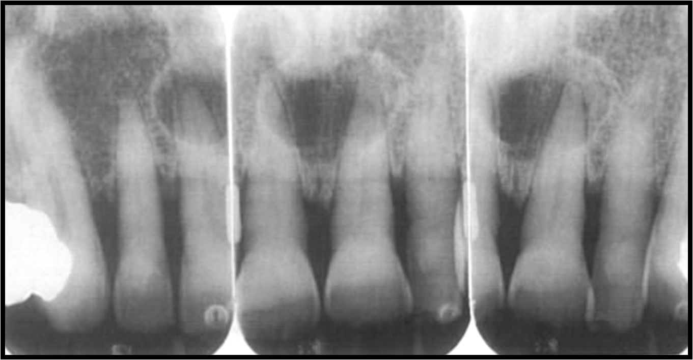
64 shows apical root lateral displacement secondary to the cystic lesion. Panoramic radiographs can be used for visualization of the mental foramen and a potential anterior looping but not for locating the mandibular incisive canal. The differential diagnosis for incisive canal cyst includes medial enlarged nasopalatine duct central giant cell granuloma central incisor root cyst.
Periapical radiograph depicting the junction of the mandibular canal and the mandibular incisive canal near the mental foramen. Contribs created this work entirely by myself color006400Contribs. This canal may also be referred to as the incisive canal.
Symmetry When evaluating radiographs first consider symmetry. Our goal is to evaluate identification of MIC by both panoramic radiograph PAN and cone-beam computed tomography CBCT. It can be single or multiple.
National Center for Biotechnology Information. Panoramic radiographs can be used for visualization of the mental foramen and a potential anterior looping but not for locating the mandibular incisive canal. An anatomical variation to be considered is the anterior looping of the mental nerve in 11 of images.
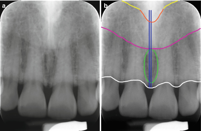
This root displacement is absent in a normal incisive canal. On periapical x-ray images the incisive foramen is located in the midline between the roots of the central incisors. It is seen on both intraoral radiographs and extraoral radiographs.
Its appearance is quite variable due to normal anatomic variation and due to the operators angulation of the x-ray beam. An anatomical variation to be considered is the anterior looping of the mental nerve in 11 of images. 150 cases with bilateral MIC were analyzed.
64 Plain film radiograph demonstrating apical root lateral displacement secondary to an incisive canal cyst. The incisive foramen also known as nasopalatine foramen or anterior palatine foramen is the oral opening of the nasopalatine canal. Incisive foramen is the opening of the incisive canal located immediately behind the maxillary central incisors.
Panoramic radiographs can be used for visualization of the mental foramen and a potential anterior looping but not for locating the mandibular incisive canal. The mean endpoint was approximately 1098 and 1026 mm anterior to the mental foramen for left and right side respectively without a. It is considered the most common non-odontogenic cyst and develops only in the midline anterior maxilla.
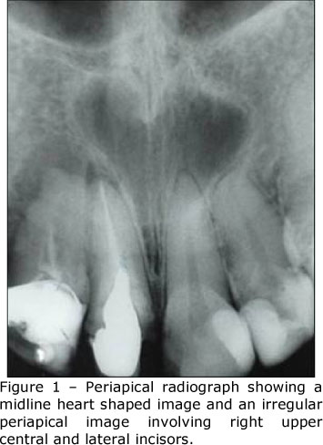
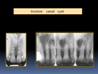
An incisive canal was identified in 15 of the images with good visibility in only 1. Results The incisive canal was found in 87 of the scans. An incisive canal cyst is a developmental cyst non neoplastic cyst arising from degeneration of nasopalatine ducts.
Incisive canal cysts are treated with complete surgical removal by a palatal approach with the palatal flap. The persistence of ductal epithelium leads to formation of cyst. 323 098 mm.
Die mittlere Breite des Foramen labiopalatal und mesiodistal betrug 312 094 mm bzw. These ducts usually regress in fetal life. RESULTS An incisive canal was identified in 93 of the cases with good visibility in 22 of the cases.
In the study by Jacobs et al 1 on 230 spiral CT scans of the mandible the incisive canal could be identified in 93 of the spiral CT scans. 30 November 2010 1346 UTC Source Original text. Popularly known as nasopalatine canal is a radiolucent tube shaped area located in between the maxillary central incisors.
The nasopalatine canal presents as a vertical radiolucent band between the roots of the maxillary central incisors superiorly to the Post topics.
Normal Anatomical Landmarks In Dental X Rays And Cbct Springerlink

Identification Of Anatomical Landmarks On A Panoramic Radiograph 1 Download Scientific Diagram

Panoramic Radiograph Image A Axial B And Oblique Sagittal C Ct Download Scientific Diagram

File Nasolabial Duct Cyst Jpg Wikipedia

Figure 2 Assessment Of The Mandibular Incisive Canal By Panoramic Radiograph And Cone Beam Computed Tomography
Intra Oral Radiographic Anatomical Landmarks

Mouth Incisive Canal Cyst Professional Radiology Outcomes

Maxillary Anterior Landmarks Intraoral Radiographic Anatomy Continuing Education Course Dentalcare Com
Radiographic Appearance Of Cysts Part 3 And Scintigraphy Intelligent Dental

Identification Of Anatomical Landmarks On A Panoramic Radiograph 1 Download Scientific Diagram
Nasopalatine Duct Cyst A Case Report Within 3 Years Follow Up

Normal Radiographic Anatomical Landmarks

Maxillary Anterior Landmarks Intraoral Radiographic Anatomy Continuing Education Course Dentalcare Com
Normal Radiographic Anatomical Landmarks

Opg Showing Incisive Foramen And Mental Foramen Download Scientific Diagram

Periapical Radiograph 1 Year After Treatment Bone And Teeth Showing Download Scientific Diagram
Figure 2 From Visibility Of Mandibular Anatomical Landmarks In Panoramic Radiography A Retrospective Study Semantic Scholar

Maxillary Anterior Landmarks Intraoral Radiographic Anatomy Continuing Education Course Dentalcare Com

Maxillary Anterior Landmarks Intraoral Radiographic Anatomy Continuing Education Course Dentalcare Com


Comments
Post a Comment